Vascular phenotyping

 with Atherosclerotic Plaques (in red), Vevo F2 System; image by FUJIFILM VisualSonics")
Vascular health and heart disease are closely related and can be evaluated by various means. Here, we have collected a battery of techniques for these purposes.

Need help?
Send an email to phenotyping@biomed.au.dk
and we will get back to you as soon as possible.
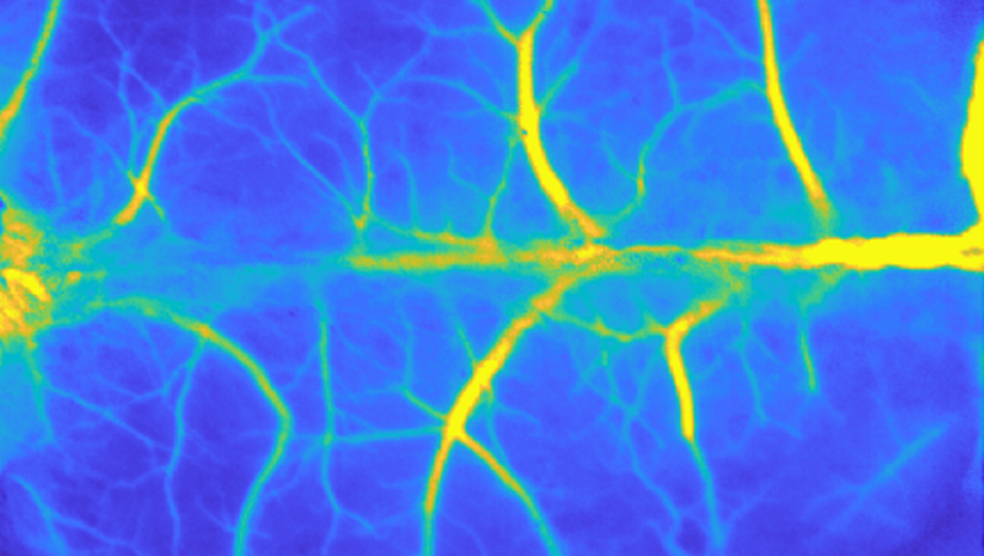
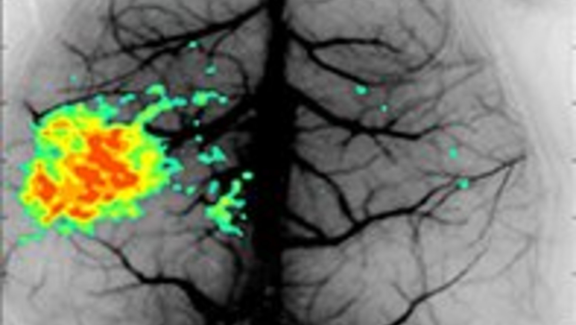
Laser Speckle Contrast Imaging (LSCI)
Applications in brief
Laser Speckle Contrast Imaging (LSCI) is a real-time imaging technique with high spatial and temporal resolution, which allows for visualization of microvascular perfusion in anaesthetized or alert animals. Speckle patterns are obtained when the biological tissue creates natural variance of backscattering of the laser signal, thus mirroring identifiable structures that can be detected using a camera. Dynamic speckles can be tracked in relation to static speckles, to track blood flow in a vessel.
Features
For investigations including:
- stroke (might require insertion of intracranial window)
- heart disease
- neurovascular flow
- arthrosis upon neurosurgery
- sunburns
- diabetic ulcers
Which species and tissues may I investigate?
The equipment is currently set up for mice in a mouse-designate location, however, the euipment can upon request be moved and set up for other species.
Training and Manual
Training and Manual
Booking
Booking
Laser Speckle Contrast Imaging
- book the equipment in the designated Teams calendar. Upon your introduction session, the Phenotyping Core Facility staff will send you an invite to the designated Teams calendar.
-
For general information on booking procedures, go to Booking
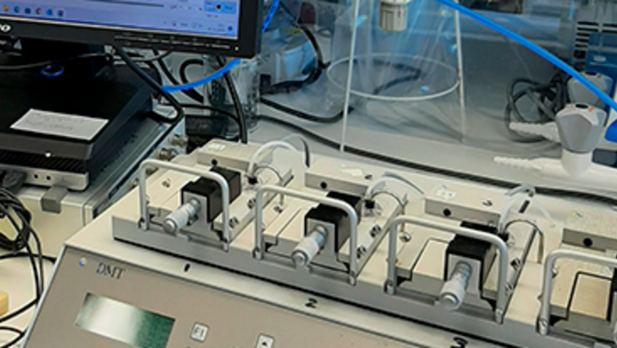
Myography
Applications in brief
The wire myograph system is used to test velocity and intensity of muscle contraction in vessels as small as 60 µm. Vessel circumference is kept constant and tension and/or relaxation in response to added compounds can be registered.
Features
For investigation of tissue including:
- aorta, basilar, coronary, mesentery and other small arteries
- veins
- lymph vessels
- tracheal rings and bladder rings
Which species may I investigate?
Tissue of any origin may be analysed in the myograph.
Training and Manual
Training and Manual

Booking
Booking
Myograph
- Book the equipment in the Outlook calendar; 1115-439 myograph
-
For general information on booking procedures, go to Booking
Ultrasound
Applications in brief
Ultrasound can be applied to visualize major arteries for subsequent functional assessment.
Features
For assesment of:
- flow velocity
- strain
Which species may I investigate?
The Vevo F2 LAZR-X system is located in the Skou Animal Facility, limiting the species to mice and rats.
Location
The Vevo F2 LAZR-X system is located in the Skou building animal facility, building 1115. Therefore, please familiarize yourself with guidelines and regulations regarding animals as well as user quarantine regulations.
Training and Manual
Training and Manual
Specifications
Specifications
| Full equipment name | Vevo F2 LAZR-X, Fujufilm Visualsonics |
| Transducer scan depths | 42 mm (UHF22x), 30 mm (UHF29x), 22 mm (UHF46x) |
| Transducer frequency and axial resolution |
|

Booking
Booking
Vevo F2 LAZR-X
- book the equipment in the Outlook calendar, 1116-K40E Vevo
-
For general information on booking procedures, go to Booking