Metabolism

Metabolic studies can be performed by various means, and equipment can often be combined and tailored to suit your exact needs - whether for long-term studies or intermittent repeated measurements.

Need help?
Send an email to phenotyping@biomed.au.dk
and we will get back to you as soon as possible.
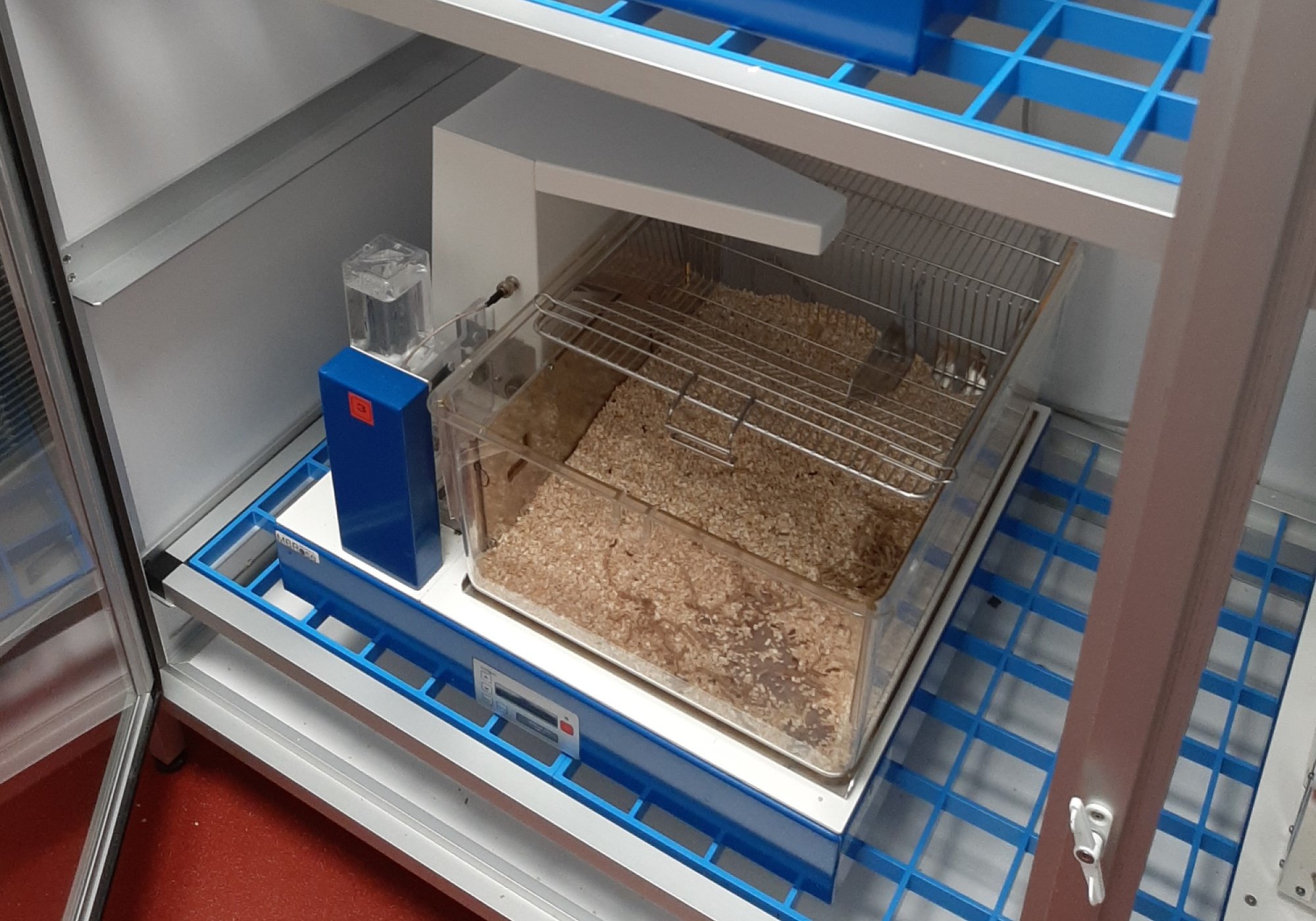
HM-2 metabolic cages
Applications in brief
HM-2 systems are advanced metabolic cages with more measurement features compared to Tecniplast metabolic cages and may be used for longer protocols, ranging from effects of drug absorption to obesity and diabetes.
Each animal is identified and monitored using its subcutaneously implanted RFID chip (requires anesthesia). The chip ensures identification of the animal as well as registration of drinking and feeding frequencies, duration, and amounts. Food and water can be manipulated, if needed.
Data is collected in the HM02Lab software, and although animals have to be weighed manually, data is logged to the same datafile/-base.
HM-2 systems can also be combined with e.g. telemetric observation of blood pressure, biopotentials and activity.
Features
For monitoring of
- food and/or water intake
- drinking and/or feeding frequencies, duration and amounts
- feces collection
- urine collection
Cages can be combined with other measurement techniques such as telemetry.
Location
HM-2 cages are normally located in the Skou building animal facility, please familiarize yourself with guidelines and regulations regarding animals as well as user quarantine regulations.
Training and Manual
Tips and Tricks
How many animals can be housed in each cage?
- 6-8 mice can be housed in each HM-2 cage.
How many cages are available?
- There are currently 4 HM-2 cages available, allowing simultaneous monitoring of up to 32 mice.
Specifications
| Full equipment name | HM-2 system, MBRose |
| Specifications | Overview on HM-2 system |
| Software | HM02Lab software |
| Chip | subcutaneous RFID chip |
Booking
HM-2 cages
- book the equipment in the Outlook calendar, 1115-K43D HM-2 cages
- for general information on booking procedures, go to Booking
Tecniplast metabolic cages
Applications in brief
Tecniplast metabolic cages can be used for registration of nutrient consumption by collecting and separating urine and faeces, e.g. for studies ranging from effects of drug absorption to obesity and diabetes.
Features
For monitoring of
- food intake
- water intake
- feces collection
- urine collection
Cages can be combined with other measurement techniques such as telemetry for observaton of e.g. blood pressure, biopotentials and activity.
Location
Tecniplast metabolic cages are normally located in the Skou building animal facility, please familiarize yourself with guidelines and regulations regarding animals as well as user quarantine regulations.
Booking
Tecniplast metabolic cages
- book the equipment by sending your request to phenotyping@biomed.au.dk
- for general information on booking procedures, go to Booking
Body composition analysis (EchoMRI)
Applications in brief
EchoMRI provides precise body composition measurements in awake animals. Body composition data include fat percentage, lean body mass, free water and total water mass, and can be obtained from mice. The scanning is fast, requiring only about 1-2 minutes, and can be repeated an unlimited number of times.
Features
For measurement of body masses of
- fat
- lean
- free water
- total water
Which species may I investigate?
- mice
Location
EchoMRI is located in the Skou building animal facility, please familiarize yourself with guidelines and regulations regarding animals as well as user quarantine regulations.
Training and Manual
Tips and Tricks
How many animals can I analyse at a time?
- 1 mouse at a time
May I use a mouse with an implanted metal device or RFID chip?
- No, EchoMRI cannot be used for mice with implanted metal devices or RFID chips. Please plan your experiments carefully, if you would like to combine EchoMRI with e.g. HM-2 metabolic monitoring.
Booking
Body composition analysis (EchoMRI)
- book the equipment in the Outlook calendar, 1115-K43B Tail cuff/MRI
- for general information on booking procedures, go to Booking